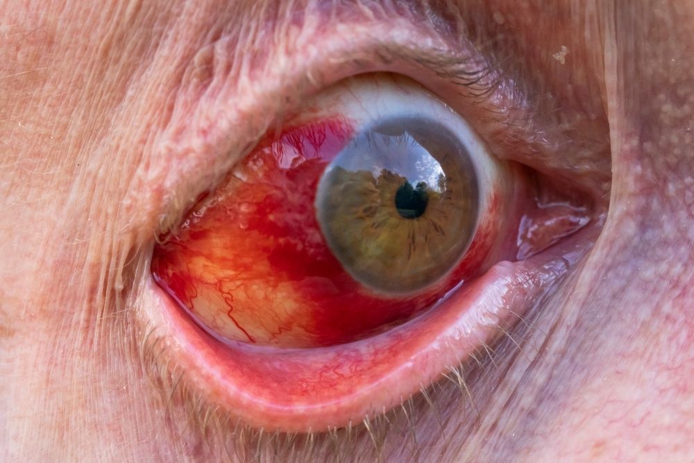
The standard diagnostic procedure, slit lamp exam, is also known as biomicroscopy. It combines a microscope with a very bright light to allow the doctor to observe the eyes in detail and determine any abnormalities.
The slit lamp exam is usually part of a comprehensive eye exam where the patient sits facing the slit lamp with their chin and forehead on a support.

How is the Slit Lamp Exam Done?
The slit lamp exam allows the doctor to clearly observe the structure of the eyes and be able to see any abnormalities. The eye doctor will have to perform an initial examination of the eyes before inserting a dye called fluorescein to make the eye exam a lot easier. This will be administered as an eye drop on a small, thin strip of paper that will touch the white part of the eye.
This will be followed by more eye drops that will dilate the pupils and enable the doctor to have a better look at the other structures of the eye. It usually takes about 20 minutes for the eye drops to take effect on the eye. Once the pupils have dilated, the doctor will repeat the eye exam but this time, a particular lens will be held close to the eye.
The Slit lamp exam is non-invasive and painless, although there may be just a brief stinging sensation during the administration of the eye drops.
A dilated pupil becomes very large which can make driving or spending time outside a little uncomfortable because the eyes become more sensitive to light. Wearing sunglasses will help during this period. Fortunately, the eye drops usually wear off after a couple of hours and the eyes will be able to return back to normal.

Interpreting the Results
This eye exam helps in the diagnosis of cataracts, corneal injuries, and scleral damage. A good range of conditions and abnormalities can also be detected through a slit lamp exam. Such abnormalities may include:
- cataracts, the opacity or clouding of the eye lens
- scleral damage
- retinal detachment
- corneal injury or disease
- retinal damage or in the blood vessels that supply it
- macular degeneration, the destruction of central vision
- disease or swelling in the middle eye layer
- glaucoma or other diseases of the optic nerve
- presence of a foreign body in the eye
- bleeding in the eye area


