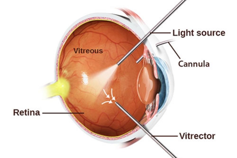
Numerous eye surgeries are being performed to improve your eye condition. Conventional laser instruments and lasers are used to perform surgeries inside the interior of the eye.
Vitreoretinal surgery refers to the surgery that occurs in the vitreous part and the retina. Surgery can restore, preserve, and enhance vision for eye conditions such as macular degeneration and diabetic retinopathy.
The referral of patients who need vitreoretinal surgery comes from a general ophthalmologist and optometrist. General ophthalmologists and other sub-specialists perform procedures involving lasers.

Procedure of a Vitrectomy Surgery
When a foreign matter enters the interior of the eye, it should be removed immediately to address the vision problems present. The vitrectomy procedure goes by removing the gel-like substance or vitreous humor in the eye.
The foreign matter present in the eye causes the shadow to appear in the retina that results in distorted or reduced vision.
A condition where blood is a foreign matter in diabetic retinopathy. Vitrectomy is used to restore the vision by removing the vitreous which is pooled with leaking blood vessels and replacing it with a clear fluid.
It starts with removing the vitreous humor and clearing the area. Injecting the saline comes next to replace the vitreous humor.
The following are the most common reasons for a vitrectomy:
- Diabetic vitreous hemorrhage
- Retinal detachment
- Epiretinal membrane
- Macular hole
- Proliferative vitreoretinopathy
- Endophthalmitis
- Foreign body removal intraocularly
General anesthesia is required in most vitrectomies, local anesthesia for certain cases.
These are the following instruments that go in the three tiny incisions made by eye surgeons for vitrectomy:
- The light pipe serves as a microscopic flashlight in the eye.
- The infusion port serves as a tube to replace the fluid with saline solution and maintain proper eye pressure.
- Vitrector is the cutting device used to remove the vitreous gel slowly. The traction is reduced when this is used to protect the delicate retina.

The Process After a Vitrectomy Surgery
The eye surgeon is the only one that knows your condition therefore, he/she can give you an idea of what will happen after the vitrectomy.
Antibiotic eye drops are usually used in the first week after the procedure and anti-inflammatory eye drops for the following weeks.
It is a must to follow the advice of your surgeon after surgery, even if this type of surgery has a very high success rate. It is rare to have potential problems such as bleeding, infection, a progression of cataract, and retinal detachment.


