Blood is carried throughout your body, including your eyes, through arteries and veins: one main artery and one main vein running through the retina of the eye. Central retinal vein occlusion occurs when the main retinal vein becomes occluded (CRVO).
Blood and fluid leak out into the retina when a vein is occluded. This fluid might cause the macula to expand, compromising your central vision. In addition, without blood circulation, nerve cells in the eye can die, and eyesight can deteriorate.
What Are the Signs and Symptoms of CRVO?

Vision loss or impaired vision in a part or all of one eye is the most prevalent sign of CRVO. It might occur quickly or gradually worsen over hours or days. However, it is possible to lose the entire vision at any time.
Floaters may be visible. In your vision, there may be black patches, lines, or squiggles. These are shadows cast by small clots of blood spilling from retinal capillaries into the vitreous.
You may experience pain and pressure in the afflicted eye in more severe cases of CRVO.
Who Is at Risk of Developing CRVO?

People over the age of 50 or with the following health issues are more likely to develop CRVO:
● diabetes
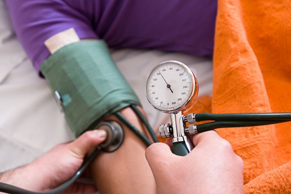
● high blood pressure
● Atherosclerosis (hardening of the arteries)
● Glaucoma
You should take the following steps to reduce your risk of CRVO:
● exercise on a regular basis
● consume a low-fat diet
● keep a healthy weight
● stop smoking
Diagnosis of Central Retinal Vein Occlusion (CRVO)

Your ophthalmologist will use eye drops to dilate (widen) your pupils and examine your retina. They will also perform an OCT scan of the retina to check for retinal edema, which causes vision loss.
Fluorescein angiography is a test that they may use. Fluorescein, a yellow dye, is injected into a vein, commonly in the arm. The dye passes through your blood vessels and into your body. As the dye flows through the vessels, a unique camera captures photographs of your retina. This test determines whether or not the retinal vein is obstructed.
People under the age of 40 who have a central retinal vein occlusion (CRVO) may be checked to see if they have a clotting or thickening problem.
What Is the Treatment for CRVO?

The primary goal of treatment is to maintain your vision. This is usually accomplished by plugging any leaky blood vessels in the retina. This helps to keep the macula from expanding any further.
Your ophthalmologist may decide to treat your CRVO with “anti-VEGF injections,” which are pharmaceutical injections into the eye. The medication can aid in the reduction of macula edema. To assist in alleviating the swelling, steroid medications may be injected into the eye.
Your ophthalmologist may perform laser eye surgery if your CRVO is severe and new abnormal blood vessels are forming in your eye. Photocoagulation of the retina is referred to as panretinal photocoagulation (PRP). The retina is burned using a laser to create microscopic burns. This reduces the risk of eye hemorrhage and keeps ocular pressure from getting too high.
It usually takes a few months after treatment to detect a difference in your eyesight. While the majority of people will notice an improvement in their eyesight, some may not.


